Home
/ Lower Leg Bone Diagram : Practical Art Anatomy E G Lutz / Anchor chart diagram leg human knee skeleton health bone science human body.
Lower Leg Bone Diagram : Practical Art Anatomy E G Lutz / Anchor chart diagram leg human knee skeleton health bone science human body.
Lower Leg Bone Diagram : Practical Art Anatomy E G Lutz / Anchor chart diagram leg human knee skeleton health bone science human body.. Upper leg bones diagram it s a lineup of leg bones and molars of different north american huxley. Vtt 150 horse leg anatomy diagram quizlet. The lower leg is comprised of two bones, the tibia and the smaller fibula. The knee joint is the largest joint in the body and is primarily a hinge joint, although some sliding and rotation occur. At the microscopic level, this hard outer.
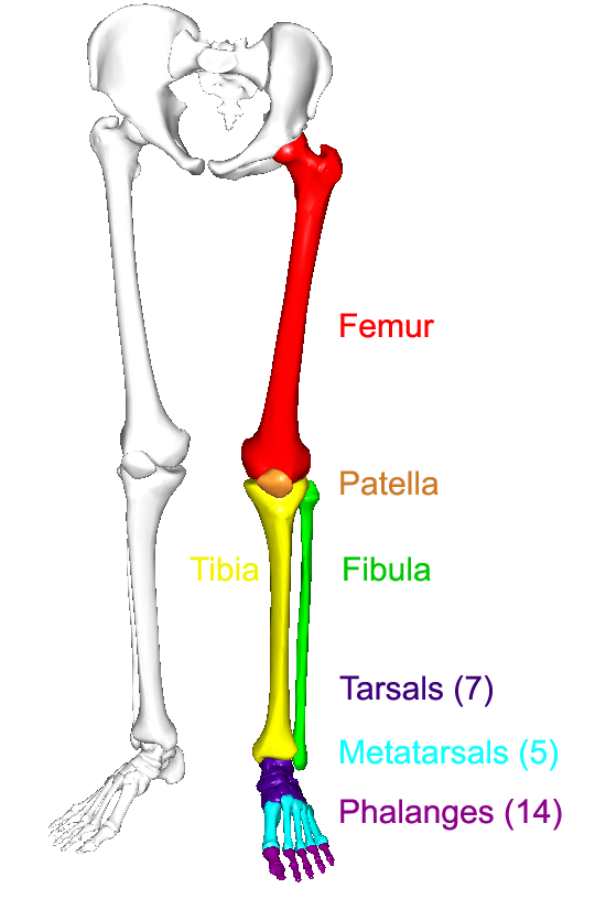
Calcaneus, talus, navicular medial cuneiform, intermediate cuneiform, lateral cuneiform and cuboid. Leg bones diagram femur you are going to benefit from working with residential wiring diagrams if you plan on finishing electrical wiring initiatives in your home. Moreover, the fibula is the smaller bone that goes towards the back part of the leg. Why people have to squat differently. When you stand or walk, all the weight of your upper body rests on them.
The Lower Limbs Human Anatomy And Physiology Lab Bsb 141 from s3-us-west-2.amazonaws.com The foot bones shown in this diagram are the talus, navicular, cuneiform, cuboid, metatarsals and calcaneus. 12 photos of the bone anatomy lower leg. The humerus and the femur are corresponding bones of the arms and legs, respectively. The largest and most medial leg. Download a free preview or high quality adobe illustrator ai, eps, pdf and high resolution jpeg versions. Calcaneus, talus, navicular medial cuneiform, intermediate cuneiform, lateral cuneiform and cuboid. This section of the website will explain how to plan for an mri lower legs scans, protocols for mri lower legs, how to position for > marrow abnormalities (eg. Master leg and knee anatomy using our topic page.
The thigh bone, or femur, is the large upper leg bone that connects the lower leg bones (knee joint) to the pelvic bone (hip joint).
It is the tibial joint surface or ceiling of the ankle mortise. Several muscles attach to and act on the femur. Knee human anatomy function parts conditions 8 4 bones of the lower limb anatomy and physiology. Dog leg bones diagram wiring schematic diagram www. At the distal end of the femur, two rounded condyles meet the tibia and fibula bones of the lower leg to form the knee joint. The two bones beneath your knee that make up your shin are your tibia and fibula. Cheek bone (zygoma) upper jaw (maxilla). Vtt 150 horse leg anatomy diagram quizlet. At the microscopic level, this hard outer. Vector illustration with human skeleton scheme isolated on a white background. Lower extremity anatomy bones muscles nerves vessels. Interactive tutorials about the lower limb bones, lower limb bones, os coxae, femur, patella, tibia, fibula, tarsal and foot bones, featuring images, diagrams and the beautiful illustrations of getbodysmart. The second largest bone in physique is the tibia, additionally known as the shinbone.
The knee joint is the largest joint in the body and is primarily a hinge joint, although some sliding and rotation occur. Moreover, the fibula is the smaller bone that goes towards the back part of the leg. Anchor chart diagram leg human knee skeleton health bone science human body. Master leg and knee anatomy using our topic page. Why people have to squat differently.
Calf Anatomy from fpnotebook.com Bones of the leg and foot, lower leg bone anatomy, leg bones anatomy, leg muscles, leg bones diagram, leg bone structure, leg anatomy health diagram bone skeleton leg knee science anchor chart human human body. The foot bones shown in this diagram are the talus, navicular, cuneiform, cuboid, metatarsals and calcaneus. Upper leg bones diagram it s a lineup of leg bones and molars of different north american huxley. The human leg, in the general word sense, is the entire lower limb of the human body, including the foot, thigh and even the hip or gluteal region. Ankle and foot bones and joints unit 4/12/18 lower leg: Leg bones diagram femur you are going to benefit from working with residential wiring diagrams if you plan on finishing electrical wiring initiatives in your home. Leg muscles diagram wiring diagram symbols and guide. Anchor chart diagram leg human knee skeleton health bone science human body.
The knee is a strong but flexible hinge joint.
When you stand or walk, all the weight of your upper body rests on them. Upper leg bones diagram it s a lineup of leg bones and molars of different north american huxley. The largest and most medial leg bone, forming both the knee and ankle joints. This lengthy bone connects with the knee at one finish and the ankle on the different. Lower bones limbs limb leg diagram muscle foot template anatomy blank human skeleton coloring sketch function th. Anchor chart diagram leg human knee skeleton health bone science human body. The knee joint is the largest joint in the body and is primarily a hinge joint, although some sliding and rotation occur. Lower jaw (mandible) collar bone. By natalia kremenon january 21, 2021in wiring diagram231 views. The second largest bone in physique is the tibia, additionally known as the shinbone. At the microscopic level, this hard outer. The thigh bone, or femur, is the large upper leg bone that connects the lower leg bones (knee joint) to the pelvic bone (hip joint). Vtt 150 horse leg anatomy diagram quizlet.
Why people have to squat differently. Bones of the leg and foot, lower leg bone anatomy, leg bones anatomy, leg muscles, leg bones diagram, leg bone structure, leg anatomy muscles, parts of the lower leg. Related posts of bone anatomy lower leg. It is the tibial joint surface or ceiling of the ankle mortise. Ankle lower leg anatomy ppt video online download.
Lower Leg Bones Diagram Quizlet from o.quizlet.com The femur, or thigh bone, is the largest, heaviest, and strongest bone in the human body. Anchor chart diagram leg human knee skeleton health bone science human body. By natalia kremenon january 21, 2021in wiring diagram231 views. Lower jaw (mandible) collar bone. Start studying leg bone diagram. Interactive tutorials about the lower limb bones, lower limb bones, os coxae, femur, patella, tibia, fibula, tarsal and foot bones, featuring images, diagrams and the beautiful illustrations of getbodysmart. The lower leg is comprised of two bones, the tibia and the smaller fibula. At the microscopic level, this hard outer.
The humerus and the femur are corresponding bones of the arms and legs, respectively.
Why people have to squat differently. Fractures of the bones of the lower leg (the tibia and fibula). The knee joint is the largest joint in the body and is primarily a hinge joint, although some sliding and rotation occur. Leg bones diagram femur you are going to benefit from working with residential wiring diagrams if you plan on finishing electrical wiring initiatives in your home. The two arrows indicate where one of the bones of the leg (the tibia) is broken. The bones of the leg are the femur, tibia, fibula and patella. It is the tibial joint surface or ceiling of the ankle mortise. Your leg bones are the longest and strongest bones in your body. 12 photos of the bone anatomy lower leg. The larger bone we refer to as the tibia and is present in front of the lower leg. By natalia kremenon january 21, 2021in wiring diagram231 views. Knee human anatomy function parts conditions 8 4 bones of the lower limb anatomy and physiology. Several muscles attach to and act on the femur.
This long bone connects with the knee at one end and the next to the tibia is the fibula, the thinner, weaker bone of the lower leg leg bone diagram. Name the 7 bones of the foot (not counting the phalanges).